
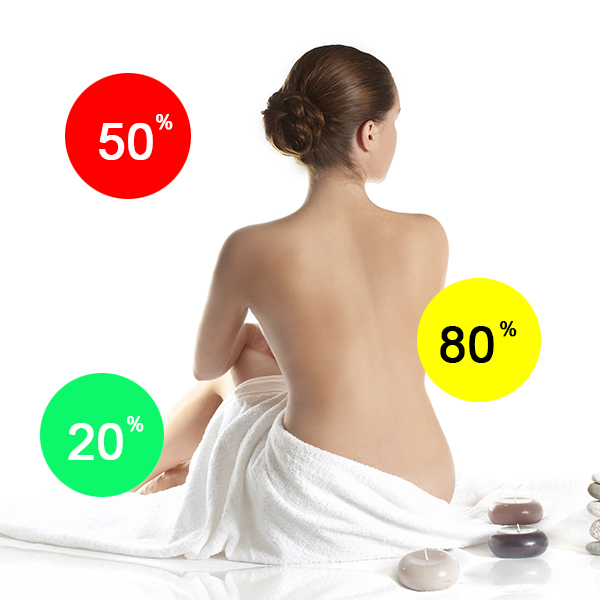

Penile extracorporeal low-intensity shock wave therapy (LIST) to the penis has recently emerged as a novel and promising modality in the treatment of erectile dysfunction (ED). LIST has angiogenic properties and stimulates neovascularization. If applied to the corpora cavernosa, LIST can improve penile blood flow and endothelial function. In a series of clinical trials, including […]
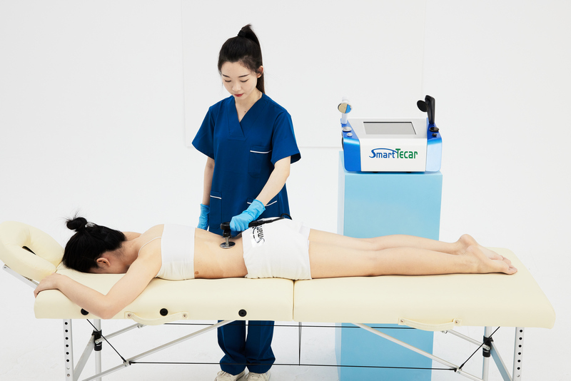
Smart tecar
Tecar's main use in physiotherapy relates to the rehabilitation of musculoskeletal and pain disorders, and sport rehabilitation.TECAR technology is non-ablative, non-invasive, using currents of high frequency, usually in the range between 300 KHz and 1Mhz.It can work in two modes of energy transfer:Capacitive mode and Resistive mode.
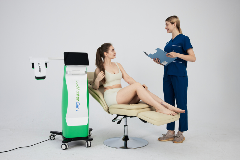
Luxmaster slim
LuxMaster Slim openes a new possibility in cellulite reduction. By using the low level laser, fat cells can be shrinked and then consumed as energy and flushed out naturally via the body's lymphatic system when combined with light exercise and sufficient water intake. It is non-invasive and no side-effects reported.

Luxmaster physio
635nm red lasers are the'therapeutic sweet spot'for treatment of pain and inflammation
405nm violet laser has been proven as an effective antiviral, antibacterial, and antifungal treatment


